Bioluminescence imaging detects and measures the bioluminescent signal in tissues using a non-invasive optical imaging system. It detects visible light from the oxidation of a substrate caused by an enzyme as a molecular reporter.
_t9tg.png)
Example
Bioluminescence imaging of primary tumor and metastases in the 4T1-GL orthotopic mammary tumor model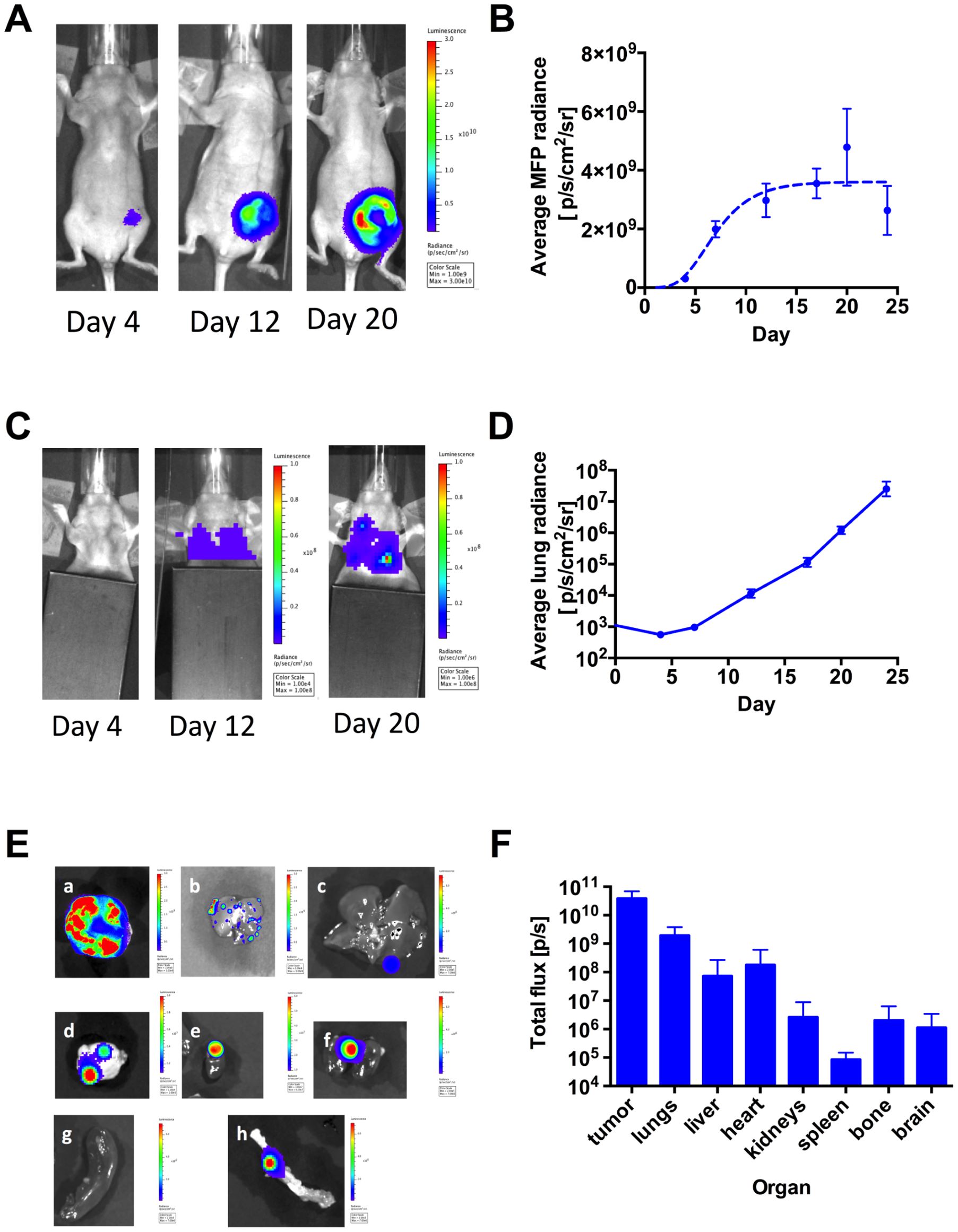
(A) Primary tumor growth in the mammary fat pad (m.f.p) as monitored using BLI, following implantation of 2×10⁷ 4T1-GL cells in the fourth left mammary fat pad (image scales in p/s/cm2/sr, n = 7 mice). (B) Corresponding quantification of BLI signal in the m.f.p area, shown as mean ± SEM and fit of the mean values to a Gompertzian tumor growth equation (dashed line, R2 = 0.86). (C) Metastases in the upper body (lung area) as monitored using BLI in the same animals (image scales in p/s/cm2/sr). (D) Corresponding quantification of the signal in the lung area, shown as mean ± SEM. (E) Metastases in excised organs on day 23
Reference
Sasportas LS, Hori SS, Pratx G, Gambhir SS. Detection and quantitation of circulating tumor cell dynamics by bioluminescence imaging in an orthotopic mammary carcinoma model. PLoS One. 2014;9(9):e105079. Published 2014 Sep 4. doi:10.1371/journal.pone.0105079